Gujarat Board GSEB Textbook Solutions Class 11 Biology Chapter 6 Anatomy of Flowering Plants Textbook Questions and Answers.
Gujarat Board Textbook Solutions Class 11 Biology Chapter 6 Anatomy of Flowering Plants
GSEB Class 11 Biology Anatomy of Flowering Plants Text Book Questions and Answers
Question 1.
State the location and function of different types of meristems.
Answer:
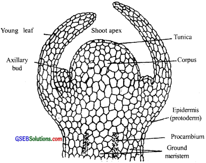
Growth in plants is largely restricted to specialized regions of active cell division called Meristems. Without meristems, normal plant growth is not possible. Plants have different kinds of meristems. The meristems which occur at the tips of roots and shoots and produce primary tissues are called apical meristems (Fig 6.1). Root apical meristem occupies the tip of a root while shoot apical meristem is present in the distant most region of the stem axis. During the formation of leaves and elongation of the stem, some ‘left behind’ cells of shoot apical meristem, constitute the axillary bud. Buds are present in the axils of leaves and are capable of forming a branch or a flower.
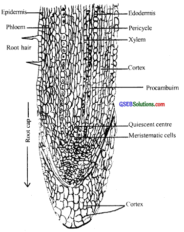
The initial apical meristems occur during the formation of an embryo. The meristem which occurs between mature tissues is known as intercalary meristem. They occur in grasses and help regenerate parts removed by the grazing herbivores. Both apical meristems and intercalary meristem are also called the primary meristem because they appear early in the life of a plant and contribute to the formation of the primary plant body.
The meristem that occurs in the mature regions of shoots and roots of many plants, particularly those that produce woody axis and appear later than primary meristem is called the lateral meristem. Generally, they are cylindrical meristems. Fascicular vascular cambium, interfascicular cambium, and cork-cambium are examples of lateral meristems.
![]()
Question 2.
Cork cambium forms tissues that form the cork. Do you agree with this statement? Explain.
Answer:
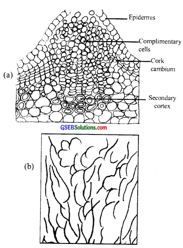
As the stem continues to increase in girth due to the activity of vascular cambium, the outer cortical and epidermis layers get broken and need to be replaced to provide new protective cell layers. Hence, another meristematic tissue called cork cambium or phellogen develops, usually in the cortex region. Phellogen is a couple of layers thick. It is made of narrow, thin-walled and nearly rectangular cells. The outer cell differentiates into the cork while inner cells differentiate into the cortex. Thus it is clear how cork cambium forms tissues that form the cork.
Question 3.
Explain the process of secondary growth in the stems of woody angiosperms with the help of schematic diagrams. What is its significance?
Answer:
The roots and stems grow in length with the help of apical meristem. It is called primary growth. Apart from growth, most di-cotyledonous plants exhibit an increase in girth. This increase is calif the secondary growth. It does not occur in monocot roots and stems. The tissues involved in secondary growth are the two lateral meristems vascular cambium and cork cambium.
The following describes the secondary growth in a dicotyledonous stem:
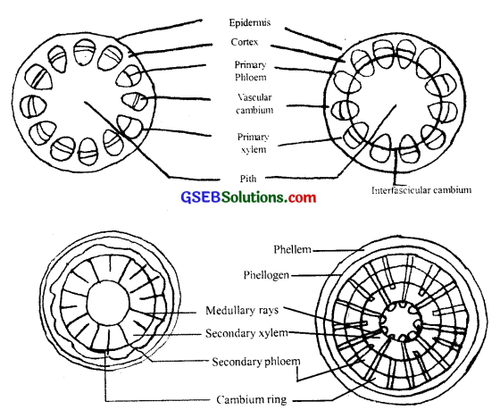
Vascular cambium The meristematic layer that is responsible for cutting off vascular tissues-xylem and phloem-is called vascular cambium. In the young stem, it is present in patches as a single layer between the xylem and phloem. Later it forms a complete ring.
Formation of cambial ring: In dicot stems, the cells of cambium present between the primary xylem and primary phloem is the intrafascicular cambium. The cells of medullary cells, adjoining this intrafascicular cambium become meristematic and form the interfascicular cambium. Thus, a continuous ring of cambium is formed.
The cambial ring becomes active and begins to cut off new’ cells, both towards the inner and the outer sides. The cells cut off towards pith, mature into secondary xylem, and the cells cut off towards periphery mature into secondary phloem. The cambium is generally more active on the inner side than on the outer. As a result, the amount of secondary xylem produced is more than the secondary phloem and soon forms a compact mass.
The primary is secondary problems get gradually crushed due to the continued formation and accumulation of secondary xylem. The primary xylem however remains more or less intact, in or around the center. At some places, the cambium forms a narrow band of parenchyma, which passes through the secondary xylem and the secondary phloem in the radial directions. These are the secondary medullary rays.
![]()
Question 4.
Draw illustrations to bring out the anatomical difference between
- Monocot root and Dicot root
- Monocot stem and Dicot stem
Answer:
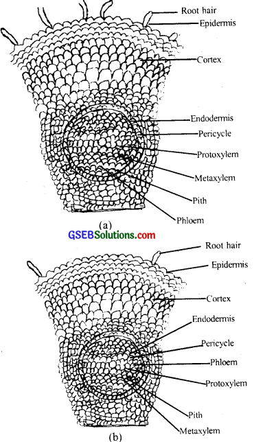
Anatomical differences between Monocot root and Dicot root
1. Monocot root:
- The cortex is very wide.
- Casparian bands are visible only in young roots. The endodermal cells later show more thickening.
- Pericycle produces lateral roots only.
- Xylem and phloem bundle 8 or more in number.
- Xylem elements are usually rounded or oval.
- Conjunctive tissue may be parenchymatous or sclerenchymatous.
- It does not produce cambium.
- A well-developed pith is present in the center of the root.
- It does not show secondary growth.
2. Dicot root:
- Cortex is comparatively narrow.
- Endodermal cells are less thick-and Casparian bands are more prominent.
- Pericycle produces lateral roots, cork, cambium, and part of the vascular cambium.
- The number of xylems and phi-OEM bundles varies from 2-6 or sometimes 8.
- Xylem elements (vessels and tracheids) are polygonal.
- Conjunctive tissue is parenchymatous.
- Conjunctive parenchyma from vascular cambium.
- Pith is either absent or very small.
- It shows secondary growth by the activity of vascular cambium and cork cambium.
Question 5.
Cut a transverse section of the young stem of a plant from your school garden and observe it under the microscope. How would you ascertain whether it is a monocot stem or a dicot stem? Given reasons.
Answer:
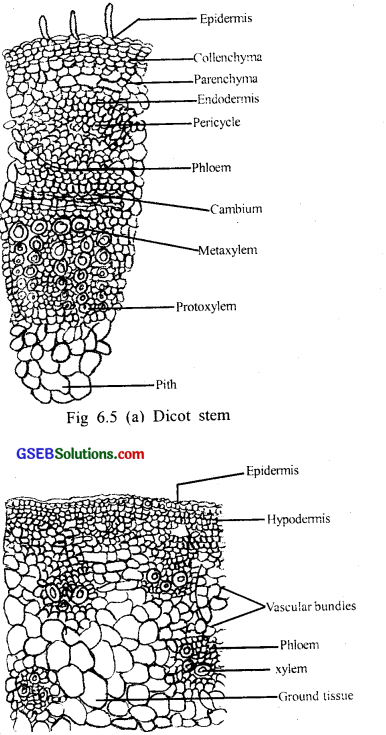
The transverse section of a typical young dicotyledonous stem shows.
(1) The epidermis, the outermost protective layer (fig 6.6 (a)) covered with a thin layer of cuticle, may bear trichomes and a few stomata.
(2) The cells arranged in multiple layers between the epidermis and pericycle constitute the cortex. It consists of three sub-zones.
- The outer hypodermis consists of a few layers of collenchymatous cells just below the epidermis, which provide mechanical strength to the young stem.
- Cortical layers below the hypodermis consist of rounded thin-walled parenchymatous cells with conspicuous intercellular spaces.
- The innermost layer of the cortex is called the endodermis. The
cells of the endodermis are rich in starch grains and the layer is also referred to as the starch’ sheath.
(3) Pericycle is present on the inner side of the endodermis and above the phloem in the form of semilunar patches of sclerenchyma.
(4) In between the vascular bundles there are a few layers of radially placed parenchymatous cells, which, constitute medullary rays. A large number of vascular bundles are arranged in a ring. The ’ring1 arrangement of vascular bundles is a characteristic of the dicot stem. Each vascular bundle is conjoint, open, and with each protoxylem. A large number of rounded, parenchymatous cells with large intercellular spaces that occupy the central portion of the stem constitute the pith.
![]()
Question 6.
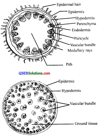
The transverse section of plant material shows the following anatomical features
1. The vascular bundles are conjoint, scattered, and surrounded by a sclerenchymatous bundle sheath,
2. Phloem parenchyma is absent. What will you identify it as?
Answer:
It is clear from the above transverse section of the plant that the plant has a monocot stem because vascular bundles are scattered In monocot stems and phloem parenchyma is absent.
Question 7.
Why are xylem and phloem called complex tissues?
Answer:
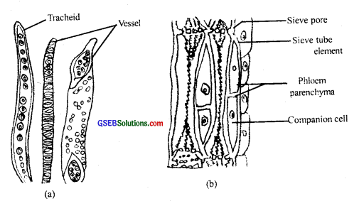
Permanent tissues having many different types of cells are called complex tissues. Because the complex tissues are made of more than one type of cell and these work together as a unit. Xylem and phloem constitute the complex tissues in plants. Xylem functions as a conducting tissue for water and minerals from roots to the stem and leaves. It also provides mechanical strength to the plant parts. It is composed of four different kinds of elements, namely, tracheids, vessels, xylem fibers, and xylem parenchyma. Gymnosperms lack vessels in their xylem. Tracheids are elongated or tube-like cells with thick and lignified walls and tapering ends.
In flowering plants, tracheids and vessels are the main water transporting elements. Xylem fibers have highly thickened walls and obliterated central lumens. Xylem parenchyma cells store food materials in the form of starch or fat and other substances like tannins. The radial conduction of water takes place by the ray parenchymatous cells, (fig 6.7 (a))
Phloem transports food materials, usually from leaves to other parts of the plant. Phloem in angiosperms is composed of sieve tube elements, companion cells, phloem parenchyma, and phloem fibers. Gymnosperms have albuminous cells and sieve cells. They lack sieve tubes and companion cells. The functions of sieve tubes are controlled by the nucleus of companion cells. The companion cells are specialized parenchymatous cells, they help in maintaining the pressure gradient in the sieve tubes, (fig 6.7 (b))
![]()
Question 8.
What is the stomatal apparatus? Explain the structure of stomata with a labeled diagram.
Answer:
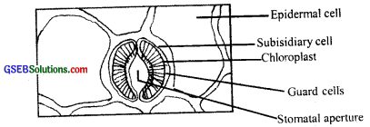
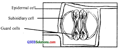
The stomatal aperture, guard cells, and the surrounding subsidiary cells are together called stomatal apparatus. Stomata are structures present in the epidermis of leaves. Stomata regulate the process of transpiration and gaseous exchange. Each stoma is composed of two bean-shaped cells known as guard cells. In grasses, the guard cells are dumbbell-shaped. The outer walls of guard cells are thin and the inner walls are highly thickened. The guard cells possess chloroplasts and regulate the opening and closing of stomata. Sometimes, a few epidermal cells, in the vicinity of the guard cells become specialized in their shape and size and are known as subsidiary cells.
Question 9.
Name the three basic tissue systems in the flowering plants. Give the tissue names under each system.
Answer:
On the basis of their structure and location, there are three types of tissue systems
- Epidermal Tissue system: Epidermis stomata
- Ground or Fundamental Tissue system: Parenchyma, sclerenchyma, and collenchyma.
- Vascular or conducting Tissue system: Phloem and Xylem.
Question 10.
How is the study of plant anatomy useful to us?
Answer:
The study of the internal structure of plants is called anatomy. Plants have cells as the basic unit, cells are organized into tissues and in turn, the tissues are organized into organs. Different organs in a plant show differences in their internal structure. Within angiosperms, the monocots and dicots are also seen to be anatomically different. Internal structures also show adaptations to diverse environments. We can very easily see the structural similarities and variations in the external morphology. the plants and we can find several similarities as well differences.
![]()
Question 11.
What is periderm? How does periderm formation take place in the dicot stems?
Answer:
As the stem continues to increase in girth due to the activity of vascular cambium, the outer cortical and epidermis layers get broken and need to be replaced to provide new protective cell layers. Hence, sooner or later, another meristematic tissue called cork cambium or phellogen develops, usually in the cortex region. Phellogen is a couple of layers thick. It is made of narrow, thin-walled, and nearly rectangular cells.
Phellogen cuts off cells on both sides. The outer cells differentiate into cork or phellem while the inner cells differentiate into secondary cortex or phelloderm. The cork is impervious to water due to suberin deposition in the cell wall. The cells of the secondary cortex are parenchymatous. Phellogen, phellem, and phelloderm are collectively known as periderm. Due to the activity of the cork cambium, pressure builds up on the remaining layers peripheral to phellogen and ultimately these layers die and slough off.
Question 12.
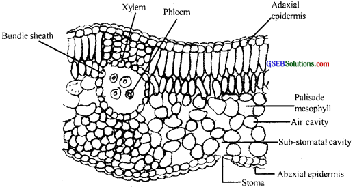
Describe the internal structure of a dorsiventral leaf with the help of labeled diagrams.
Answer:
Mesophyll, which possesses chloroplasts and carries out photosynthesis, is made up of parenchyma. It has two types of cells – the palisade parenchyma and the spongy parenchyma. The adaxially placed palisade parenchyma is made up of elongated cells, which are arranged vertically and parallel to each other. The oval or round and loosely arranged spongy parenchyma is situated below the palisade cells and exter ds to the lower epidermis.
There are numerous large spaces and air cavities between these cells. The vascular system includes vascular bundles, which can be seen in the veins and the midrib. The size of the vascular bundles are dependent on the size of the veins. The veins vary in thickness in the reticulate venation of the dicot leaves. The vascular bundles are surrounded by a layer of thick-walled bundle sheath cells.