Gujarat Board GSEB Textbook Solutions Class 11 Biology Chapter 20 Locomotion and Movement Textbook Questions and Answers.
Gujarat Board Textbook Solutions Class 11 Biology Chapter 20 Locomotion and Movement
GSEB Class 11 Biology Locomotion and Movement Text Book Questions and Answers
Question 1.
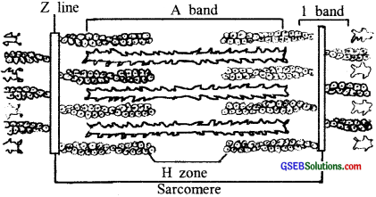
Draw the diagram of a sarcomere of skeletal muscle showing different regions.

Question 2.
Define sliding filament theory of muscle contraction.
Answer:
The mechanism of muscle contraction is best explained by the sliding filament theory which states that contraction of a muscle fiber takes place by the sliding of the thin filaments over the thick filaments.
![]()
Question 3.
Describe the important steps in muscle contraction.
Answer:
‘Muscle Contraction is initiated by a signal sent by the central nervous system (CNS) via a motor neuron. A motor neuron along with the muscle fibers connected to it constitutes a motor unit. The junction between a motor neuron and the sarcolemma of the muscle fiber is called the neuromuscular junction or motor-end plate. A neural signal reaching this junction releases a neurotransmitter which generates an action potential in the sarcolemma. This spreads through the muscle fibre and causes the release of calcium ions into the sarcoplasm.
Increase in Ca++ level leads to the binding of calcium with a subunit of troponin on actin filaments and thereby removes the masking of active sites for myosin. Utilising the energy from ATP hydrolysis, the myosin head now binds to the exposed active sites on actin filaments towards the centre of ‘A’ band. The Z’ line attached to these actins are also pulled inwards thereby causing a shortening of the sarcomere, i.e contraction. During contraction, the T bands get reduced, whereas the ‘A’ bands retain the length.
The ATP is again hydrolysed by the myosin head and the cycle of cross-bridge formation and breakage is repeated causing further sliding. The process continues till the Ca++ ions are pumped back to the sarcoplasmic Cisternae resulting in the masking of actin filaments. This causes the return of Z lines back to their original position, i.e. relaxation.
![]()
Question 4.
Write true or false: If false change the statement so that it is true.
- Actin is present in thin filament
- H-zone of striated muscle fiber represents both thick and thin filaments.
- The human skeleton has 206 bones.
- There are 11 pairs of ribs in man.
- The sternum is present on the ventral side of the body.
Answer:
- True
- True
- True
- False. There are 12 pairs of ribs in man.
- True
Question 5.
Write the difference between:
- Actin and myosin
- Red and white muscles
- Pectoral and Pelvic girdle
Answer:
(1) Differences between Actin and Myosin:
Actin:
- These are thin filaments.
- Actin has low molecular weight filamentous protein.
- It occurs in two forms, monomeric G-actin and polymeric F-actin.
- The thin filaments also contain the contractile protein, called tropomyosin.
- It is a rod-shaped fibrous protein.
Myosin:
- These are thick filaments.
- Myosin has high molecular-weight, small globular proteins.
- It masks the active sites of F actin.
- Each myosin molecule has two components, a tail, and ahead.
- It is a globular protein.
(2) Differences between Red and White muscles:
Red muscles:
- They are smaller in diameter.
- Mitochondria are more in number.
- Blood capillaries are more.
- The sarcoplasmic reticulum is less.
- They contain a very high amount of myoglobin.
White muscles:
- They are bigger in diameter.
- Mitochondrial are less in number.
- Blood capillaries are less.
- The sarcoplasmic reticulum is more.
- They contain a very low amount of myoglobin.
(3) Differences between the Petrol and Pelvic girdle:
Pectoral girdle:
- Petrol bones help in the articulation of the upper and the lower limbs respectively with the axial skeleton.
- The petrol girdle is formed of two halves.
- Each half of the pectoral girdle consists of a clavicle and a scapula.
- The scapula is a large triangular flat bone situated in the post¬erior part of the thorax between the second and the seventh ribs.
- The posterior, flat, triangular body of the scapula has a slightly elevated ride called the spine which projects as a flat, expanded process called the acromion.
- The clavicle articulates with this. Below the acromion is a depression called the glenoid cavity which articulates with the head of the humerus to form the shoulder joint.
- Each clavicle is a long slender bone with two curvatures. This bone is commonly called the collarbone.
Pelvic girdle:
- The pelvic girdle also helps in the articulation of the upper and the lower limbs respectively with the axial skeleton.
- The pelvic girdle consists of two coxal bones.
- Each coxal bone is formed by the fusion of three bones ilium, ischium, and pubis.
- At the point of fusion of the above bones is a cavity called the acetabulum to which the thigh bone articulates.
- Anteriorly, the two halves of the pelvic girdle meet to form the pubic symphysis containing fibrous cartilage.
![]()
Question 6.
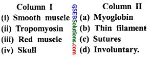
Match column I with column II:

Answer:
- d
- b
- a
- c
Question 7.
What are the different types of movements exhibited by the cells of the human body?
Answer:
Cells of the human body exhibit three main types of movements, namely, amoeboid, ciliary and muscular.Some specialised cells in our body like macrophages and leucocytes in blood exhibit amoeboid movement. It is affected by pseudopodia formed by the streaming of protoplasm.
Ciliary movement occurs in most of our internal tubular organs which are lined by ciliated epithelium. The movement of cilia in the trachea help in removing dust particles and some foreign substances. Passage of ova through the female reproductive tract is also facilitated by the ciliary movement. Movement of our limbs, jaws, tongue etc. requires muscular movement. The contractile property of muscles is effectively used for locomotion and other movements by human beings.
![]()
Question 8.
How do you distinguish between a skeletal muscle and a cardie muscle?
Answer:
| Skeletal Muscle | Cardiac Muscle |
| (i) Known as striped muscle | (i) Known as heart muscle. |
| (ii) Voluntary in function | (ii) Involuntary in function |
| (iii) Several nuclei, peripherally placed | (iii) One or more nuclei centrally placed. |
| (iv) Attached to the skeleton in trunk, limbs, and head | (iv) Found only in the walls of heart chambers |
| (v) Intercalated disc absent. | (v) Intercalated disc present. |
| (vi) Powerful and rapid contraction seen | (vi) Rhythmical contraction and relaxation seen |
Question 9.
Name the type of joint between the followings:
- Atlas/ Axis
- Carpal/ metacarpal of the thumb
- between phalanges
- Femur/acetabulum
- between cranial bones
- between pubic bones in the pelvic girdle.
Answer:
- Pivot joint
- Ball and socket
- Saddle joint
- Fibrous joint
- Gliding joint
- Ball and socket
![]()
Question 10.
Fill in the blank spaces:
Answer:
- All mammals (except a few) have seven cervical vertebrae.
- The number of phalanges in each limb of a human is 14.
- The thin filament of a myofibril contains 2 ‘F’ actins and two other proteins namely Tropomyosin and Troponin.
- In a muscle fiber, Ca++ is stored in Sarcoplasm.
- 11th and 12th pairs of ribs are called floating ribs.
- The human cranium is made of 8 bones.